The x ray diffraction experiment requires an x ray source the sample under investigation and a detector to pick up the diffracted x rays. It provides information on structures phases preferred crystal orientations texture and other structural parameters such as average grain size crystallinity strain and crystal defects.
 Usgs Ofr01 041 X Ray Diffraction Primer
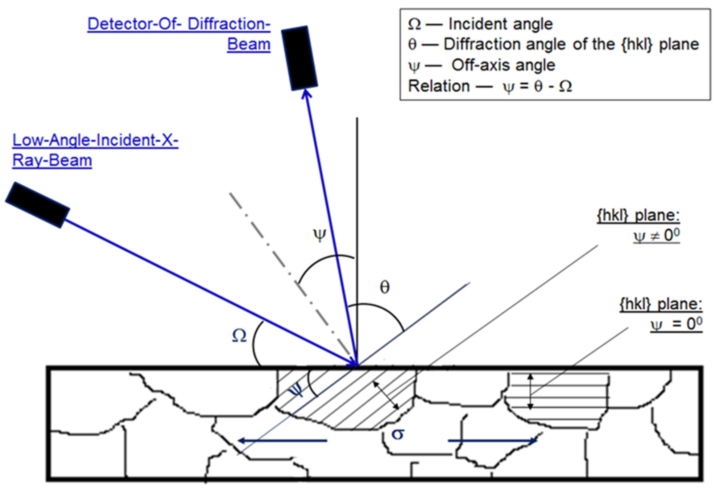
The powder diffractometers typically use the bragg brentano geometry w 2 the incident angle w is defined between the x ray source and the sample.

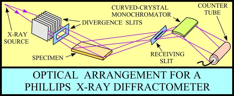
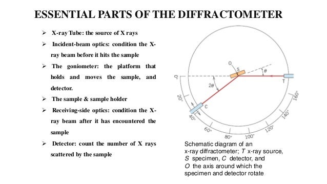
X ray diffractometer schematic diagram. Divergent slits located between the x ray source and the specimen and divergent slits located between the specimen and the detector limit scattered non diffracted radiation reduce background noise and collimate the radiation. Figure 1 is a schematic diagram of a powder x ray diffractometer. Schematic diagram of youngs double slit experiment.
The cathode part of x ray tube generated electrons under electric current. Synchrotron radiation is a brighter source and as a result can increase the resolution. X ray tube equipped with filter is commonly used in laboratory diffractometer.
Schematic diagram of x ray tube. A lead shield is use to collect the x rays after striking on film. The diffracted angle 2 is defined between the incident beam and the.
An x ray diffractometer is equipped with a position sensitive detector and a collimator preceding the detector. X ray tube is a common source of x rays. Source produces high voltage supply which are rectified by rectifier and applied to anode of the x ray tube.
X ray diffraction xrd is a powerful nondestructive technique for characterizing crystalline materials. Possible x ray sources are x ray tube synchrotron radiation and cyclotron radiation. Typical view of x ray diffractometer.
The article will provide basic details on the component parts of the x ray diffractometer. Figure 32 x ray tube. Constructive and destructive interference of waves constructive interference in phase destructive interference out of phase.
The basic geometry of an x ray diffractometer involves a source of monochromatic radiation and an x ray detector situated on the circumference of a graduated circle centered on the powder specimen. It comprises of an evacuated tube which contains a copper block anode bearing a metal target made of any of the metals such as molybdenum tungsten copper. Schematic diagram of x ray diffraction.
Slits s 1 and s 2. Diffraction occurs only when the distance travelled by the parallel x rays are an integer of the wavelength. In phase means that the peak of one wave matches the peak of the following wave.
Smallest building block c a b a g unit cell a cscl b d1 lattice d2 a crystal consists of a periodic arrangement of the unit cell into a lattice. The x rays passed from the desired region of the patient body are made to strike on the film where they produce an image of the body soft and hard parts. The lamellae of the collimator are radially aligned to the specimen which is arranged in the center of a measurement circle along which the detector and collimator move during a measurement.
X rays diffracted in phase will give a signal.
 Energy Dispersive X Ray Diffraction Wikipedia
Powder X Ray Diffraction
Gale Academic Onefile Document The Optics And Alignment Of The
 X Ray Diffraction Bruker D8 Discover Institute For Nuclear And
 Coatings Free Full Text Uncertainty Of The X Ray Diffraction
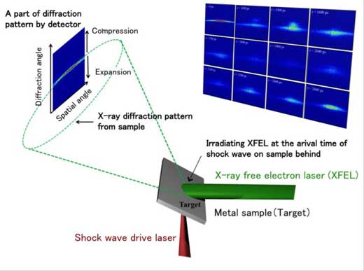 What Happens When Materials Collide Observing Fracture In
 X Ray Diffractometer And Its Various Component Parts For X Ray Studies
 Crystallography Iii X Ray Diffraction
 X Ray Diffraction Xrd Techniques For Materials Characterization
Https Xray Chem Tamu Edu Pdf Notes Intro2xrd Pdf
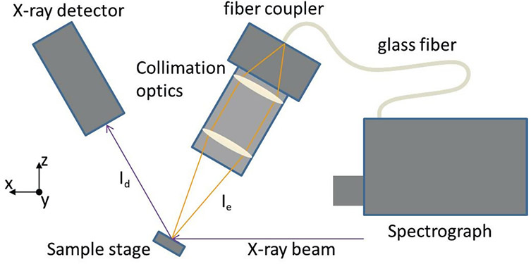 X Ray Excited Optical Luminescence With High Resolution X Ray
 7 3 X Ray Crystallography Chemistry Libretexts
Xrd Instrument Images
 Schematics Of X Ray Diffractometer 12 Download Scientific Diagram
 X Ray Diffraction Technique Xrd
High Resolution X Ray Diffraction Analyses Of La Sr Mno Sub 3
 X Ray Diffraction Xrd Schematic Diagram X Ray Diffraction Xrd
 X Ray Diffraction Xrd Ppt Video Online Download
No comments:
Post a Comment